3401 Civic Center Blvd
Philadelphia, PA, 19104
(215) 590-4545
BIOMEDICAL VISUAL COMMUNICATION
Your Custom Text Here

Your Custom Text Here
As technology advances, so too does the ability to develop new and cutting edge surgical techniques which aid in the speed of recovery, aid in safety, and provide outcomes that were before unatainable.
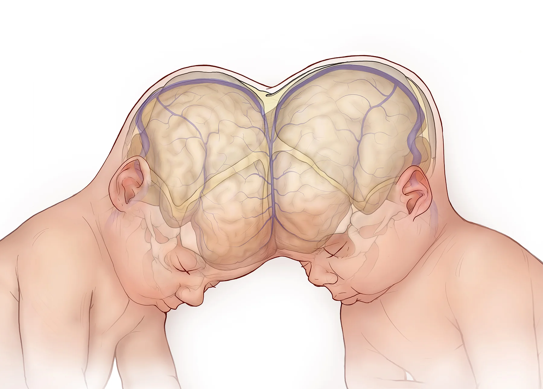
When conjoined twins are joined at the head they are referred to as caraniopagus. This series of illustrations was created to help describe the specifics of the the twins anatomy and how they were successfully separated.
This illustration was incorporated into the press release as part of the artificial womb research at The Children's Hospital of Philadelphia. The illustration posted on a vast array of news organizations around the world. This image was pulled from home page of National Public Radio (NPR).
When a fetus is diagnosed with a myelomeningocele (MMC), or more commonly known as Spina Bifida there is now a procedure which can give some families a chance at improved outcomes and a potentially higher quality of life for the child. This procedure occurs when the fetus is still developing in the the womb and requires the highest level of surgical technique. This series of illustrations created for a fetal surgical atlas, and is primarily drawn in graphite.
This axial cross section shows the defect prior to surgery, the creation of the myofacial flap, and the final sutured closure.
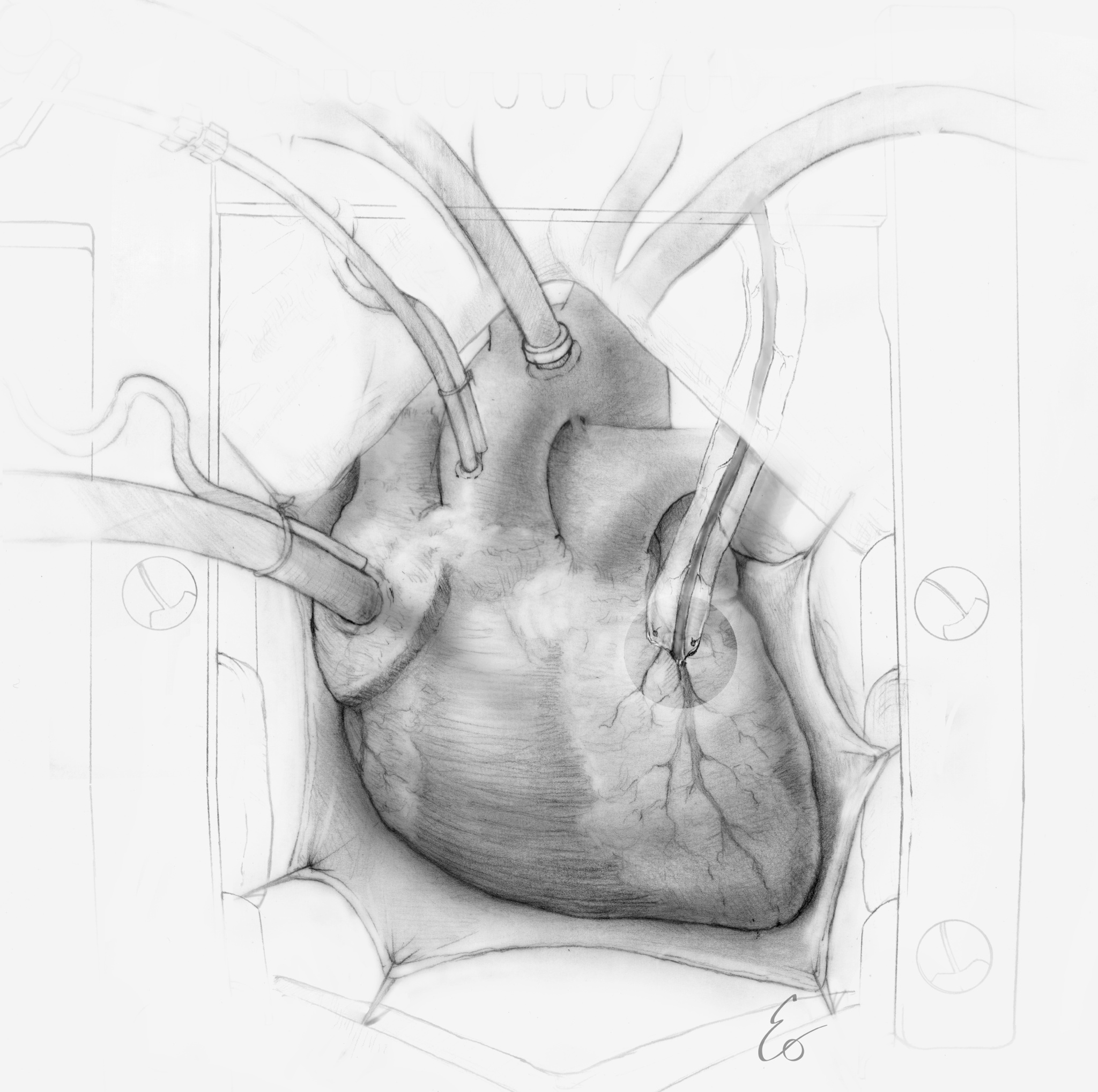
When the coronary arteries of the heart become clogged, it then may be time for a bypass procedure. This graphite illustration shows the surgical view of the procedure including the grafted artery to the heart, and the cannulation, which carries blood to and from the oxigenator.
The VEPTR device is used to treat thoracic insufficiency syndrome (TIS). TIS prevents normal development and function of the lungs. The VEPTR device is used to separate and expand the ribs of the ribcage to create volume and in the process the device can additionally help straighten the spine in cases of extreme scoliosis.
The key to finding avenues for clinical and medical research to make an impact is to make certain the science can be understood. The studio takes complex concepts and large data sets and distills them into visuals easily understood by a diversity of audiences.
This composition describes the concept of how gene editing is studied to further advance the treatment of genetic disorders while the patient is in the womb.
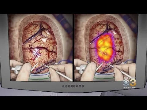
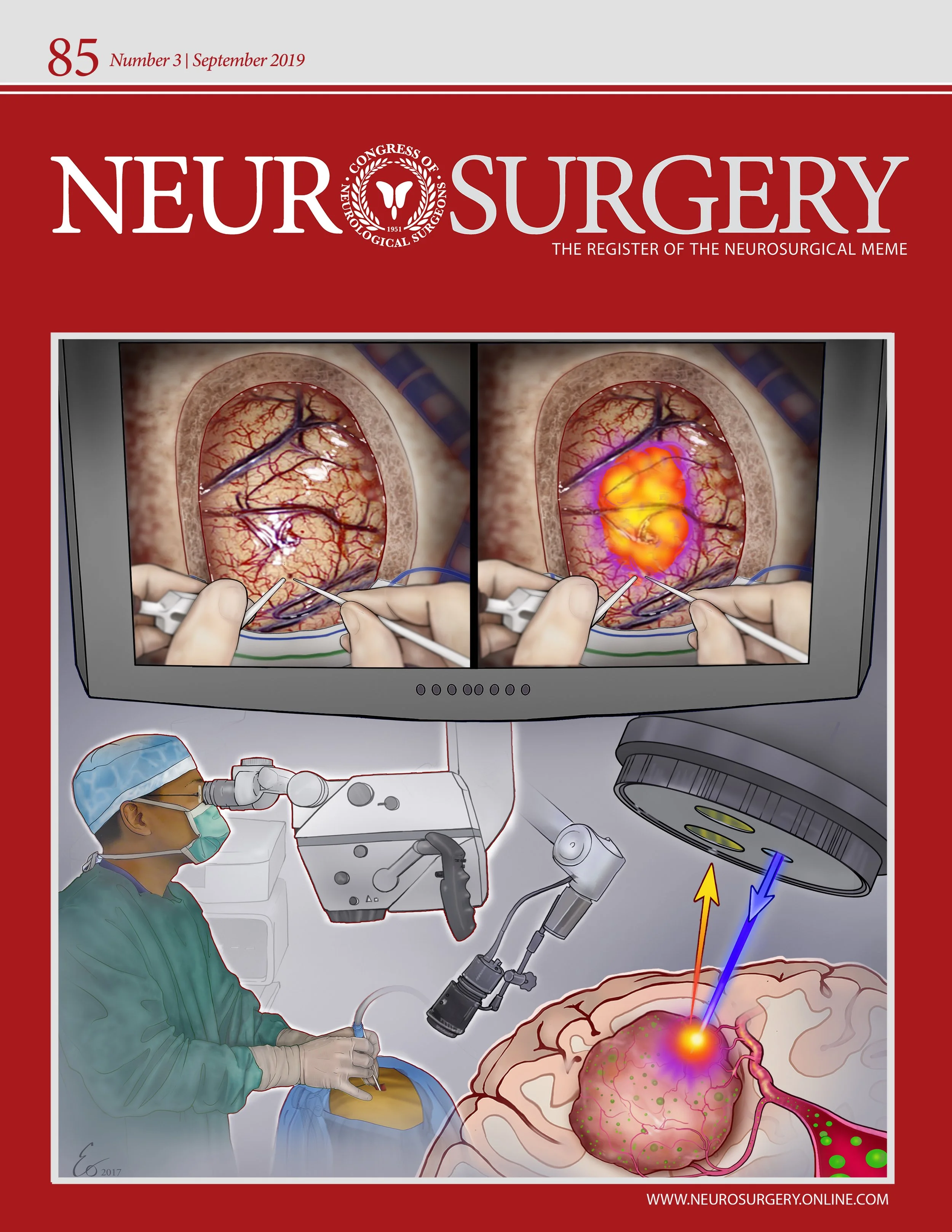
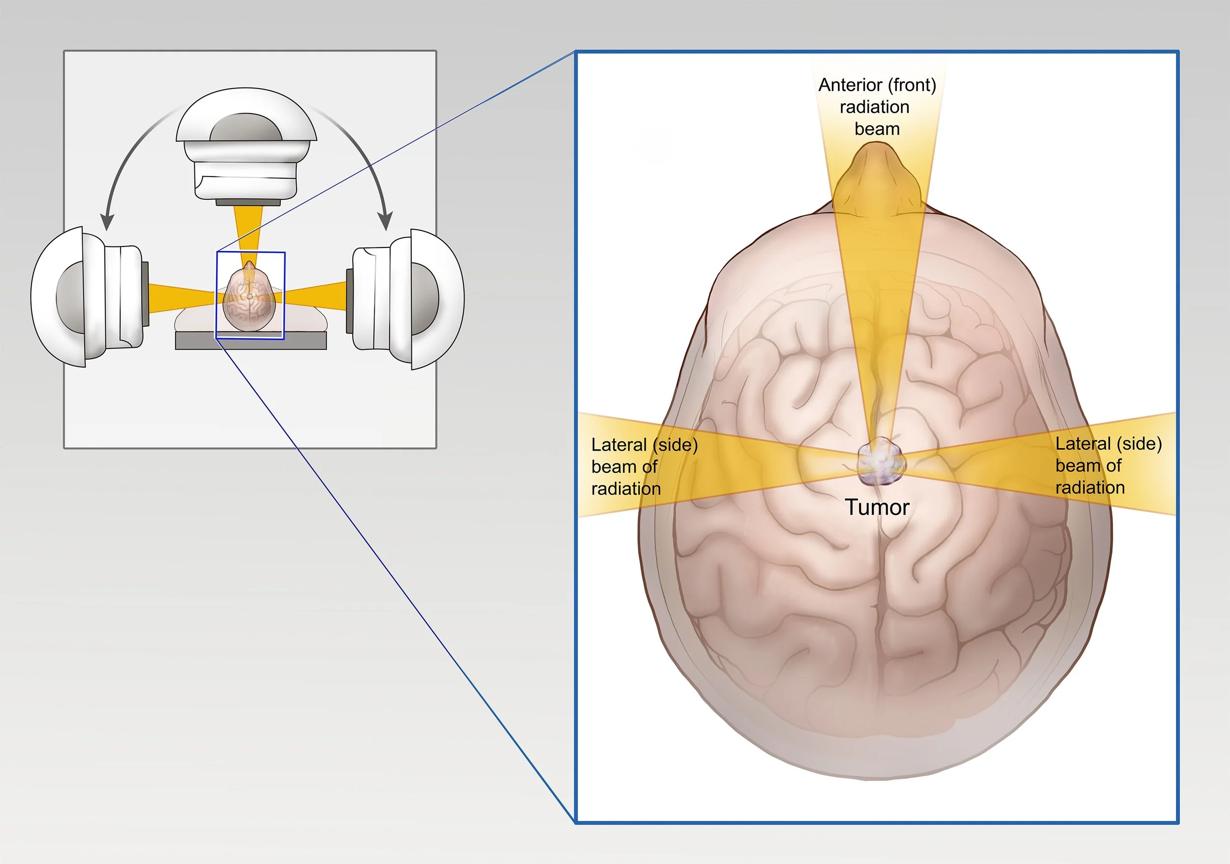
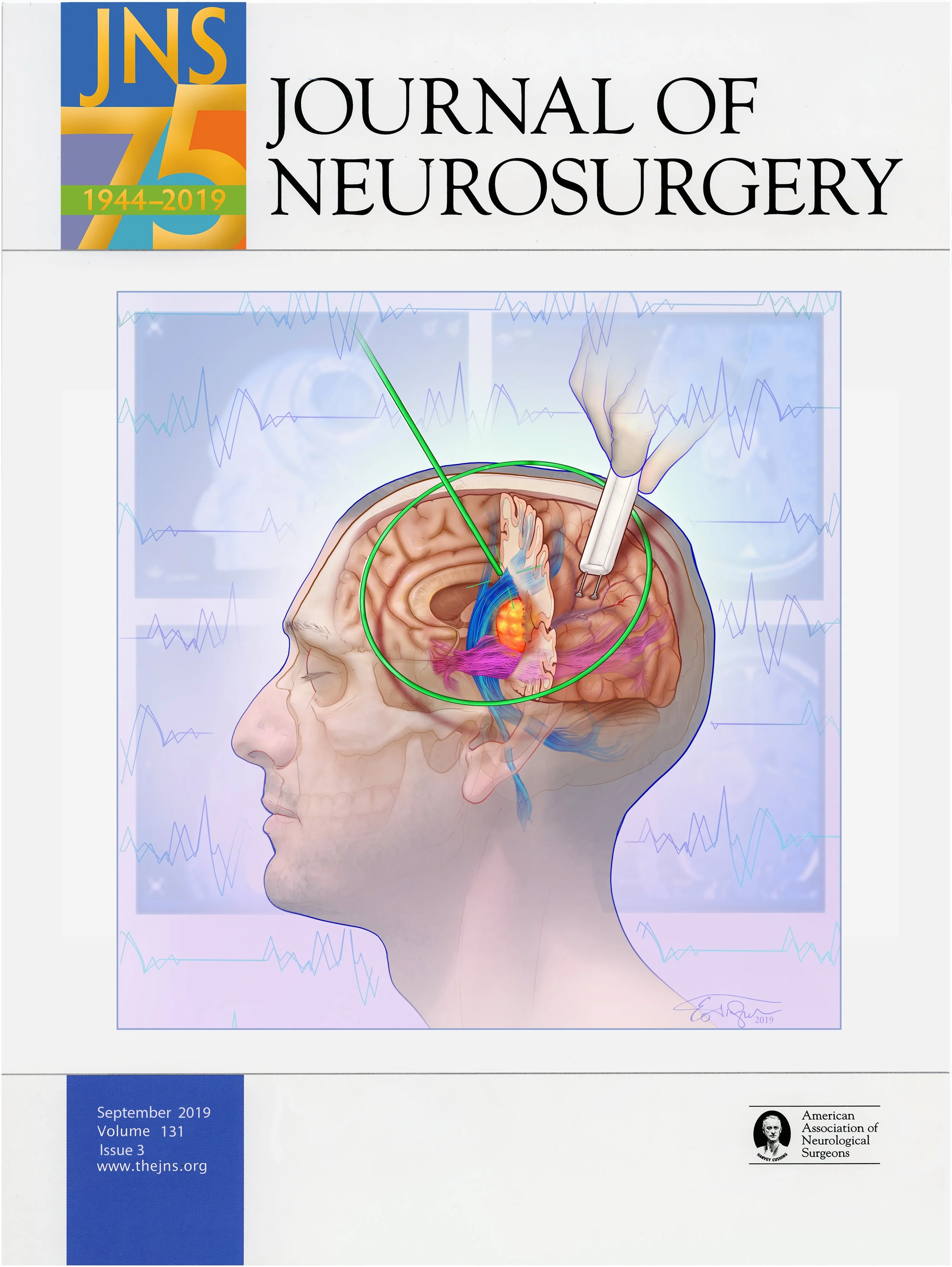
The illustrations developed for Dr. John Lee and journal cover for Neurosurgery help describe the emerging science of brain tumor removeal.
The cover illustration shows a reimagined concept of how blood in the fetal heart "sculpts" the aortic valve.
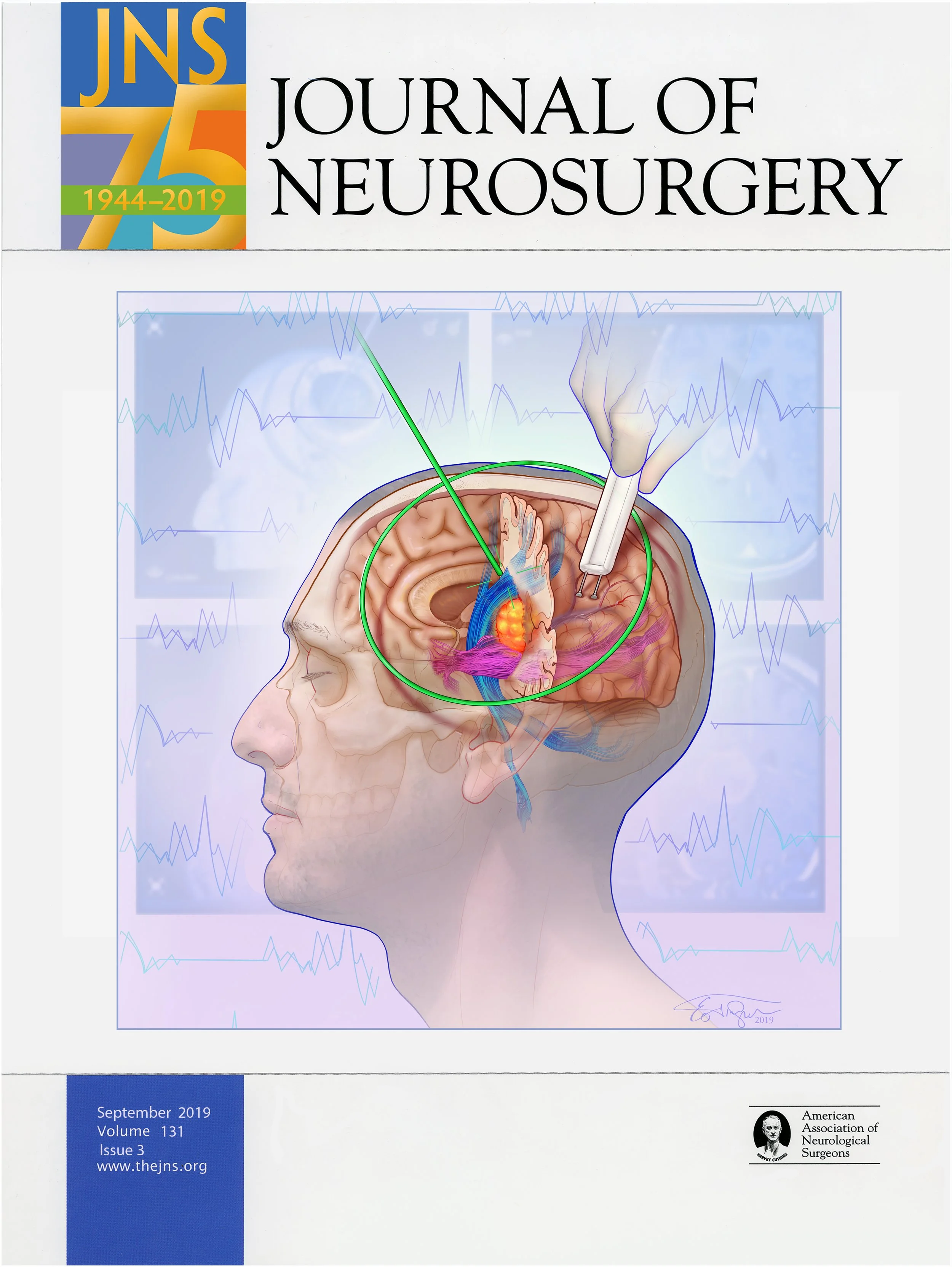
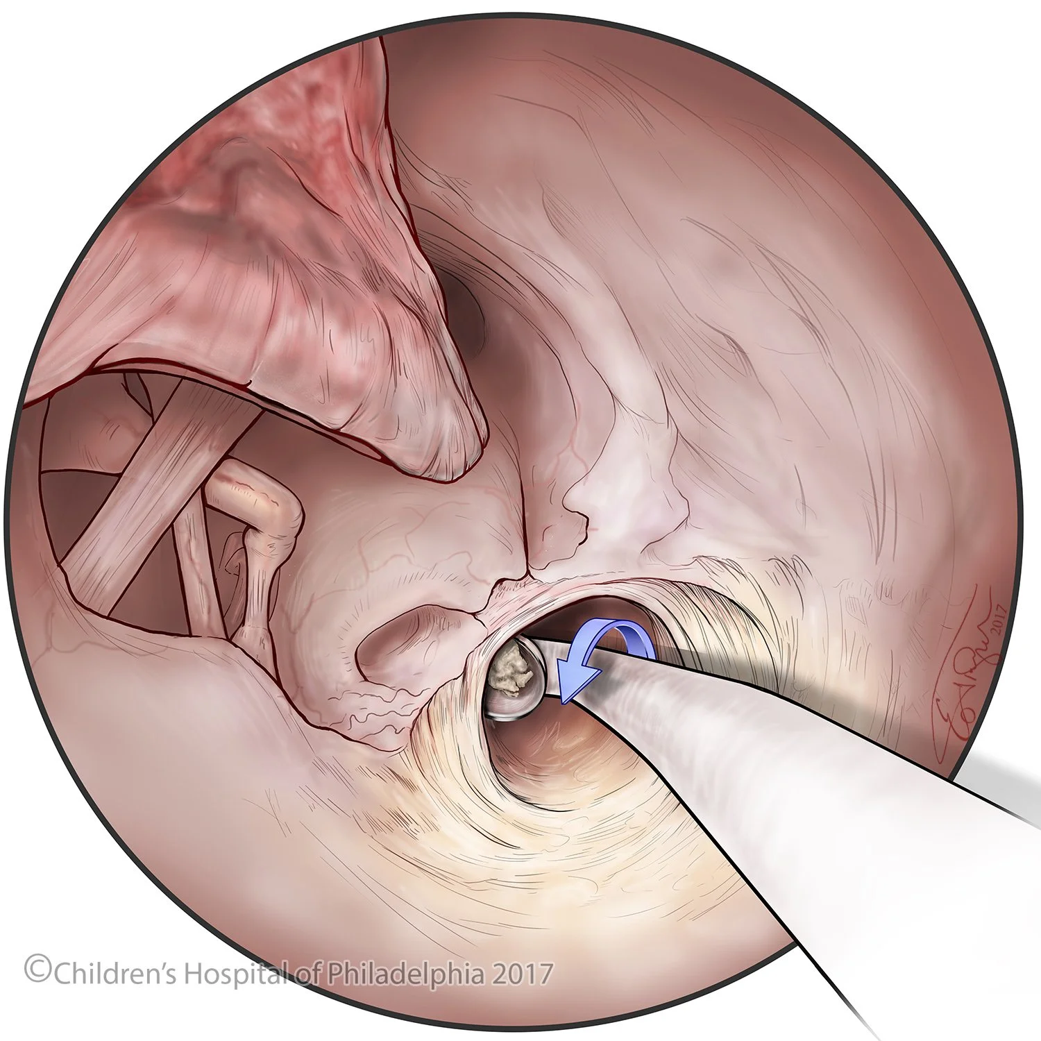
The cover composition for the May 2017 issue of the Journal of Neurosurgery (JNS) shows the key elements of a novel endoscopic approach for treating trigeminal neuralgia. The illustration was created in collaboration with Dr. John Lee at the Hospital of the University of Pennsylvania.
Stylus is the publication at the University of Pennsylvania which covers the crossroads of medicine, art, and literature. This illustration was awarded the cover in 2015.
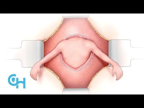
In July 2015 The Children's Hospital of Philadelphia performed the first ever bilateral hand transplant on a child. I was fortunate enough to illustrate the procedure. The illustrations were used to educate the public about the steps of the procedure and were used as part of the hospital's press release.
This video shows the ground breaking surgery to remove a tumor from lining of the heart of a fetus in the womb.
This is Zion's story. It chronicles his journey of getting two new hands. My illustrations are used to bring clarity to the surgical procedure.
A pre surgery view of the patient and his arms.
After the veins and nerves are reattached, over a dozen muscles and tendons are connected.
After a surgery that lasted more than half a day, the patient has new hands.
The complexity of the body' structure and its function is best described when strong clear visuals are accompany written text. The following examples focuses on such examples.
This is illustration shows the section of the knee joint that sits atop the tibia bone.
The blood supply to the neck of the femur has been poorly understood for years. This illustration tracks the the course of the arteries and highlight their relationship to the muscles of the hip.
The complexity of the human knee is only matched by its beauty.
The two illustrations shown here provide an anterior (frontal) view and an axial (top) view of the knee joint. Particular attention is given to the ligaments and tendons of the joint.
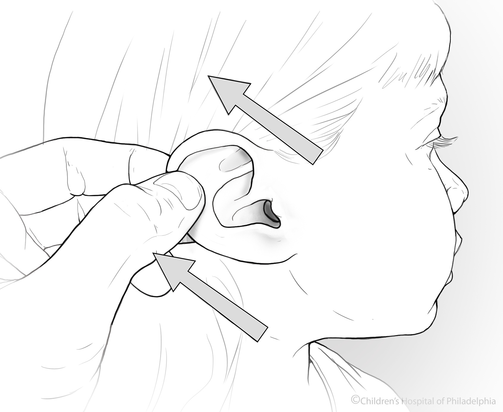
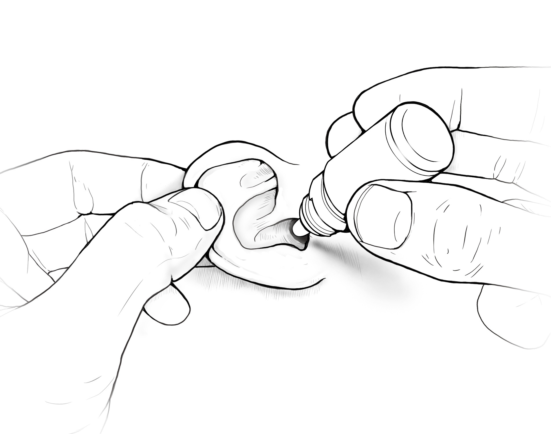
Once a child comes home from the hospital the family becomes the primary care givers tasked with administering medications, maintaining medical devices, and monitoring the health of the child. Education is required to get parents and patients up to speed in terms of knowledge and techniques.
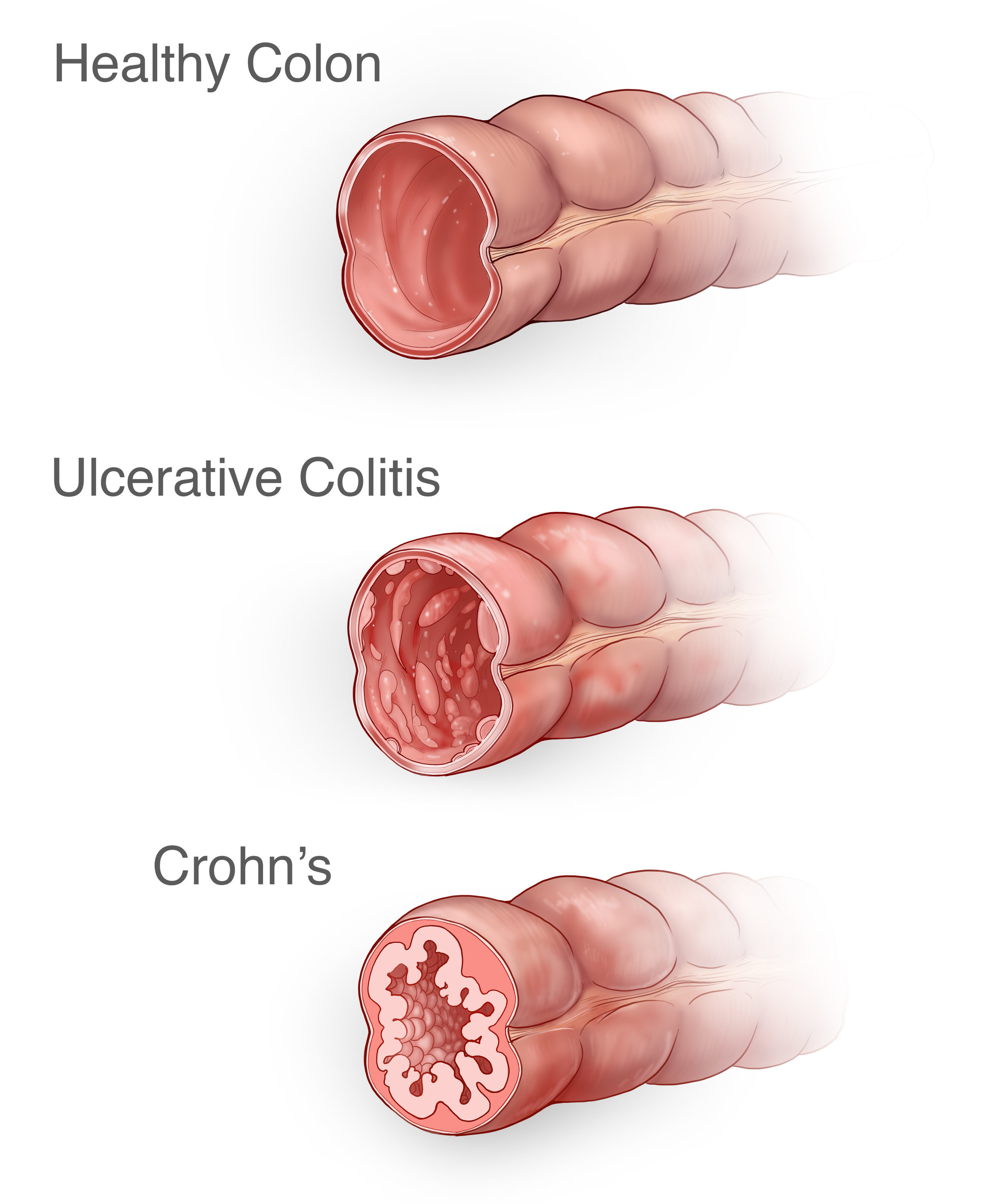
Inflammatory Bowel Disease or IBD is a general term for chronic conditions of the intestines stemming from inflammation and ulcerations. The two most common IBD conditions are Crohn's and Uclerative Colitis.
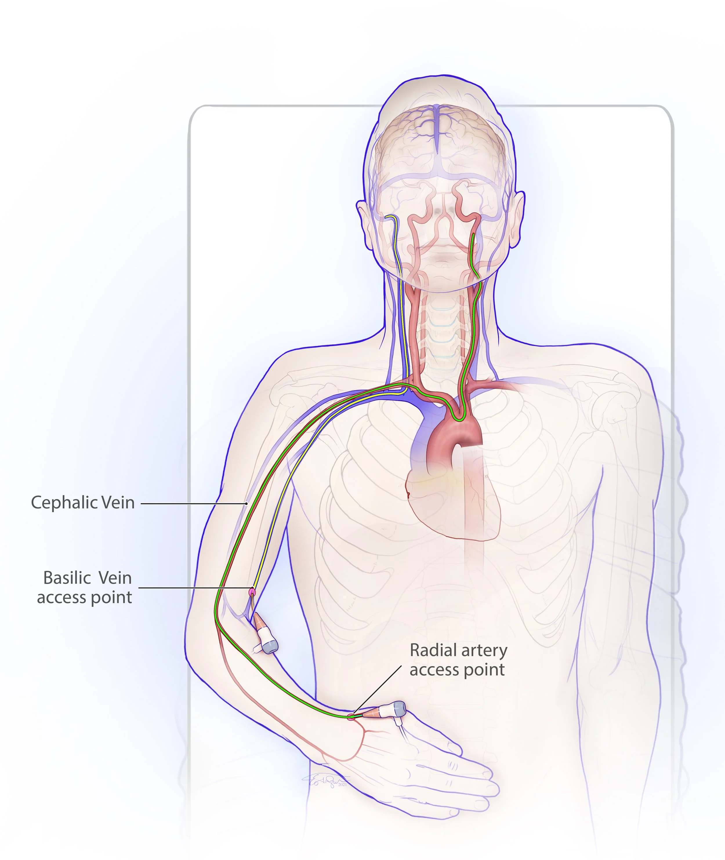
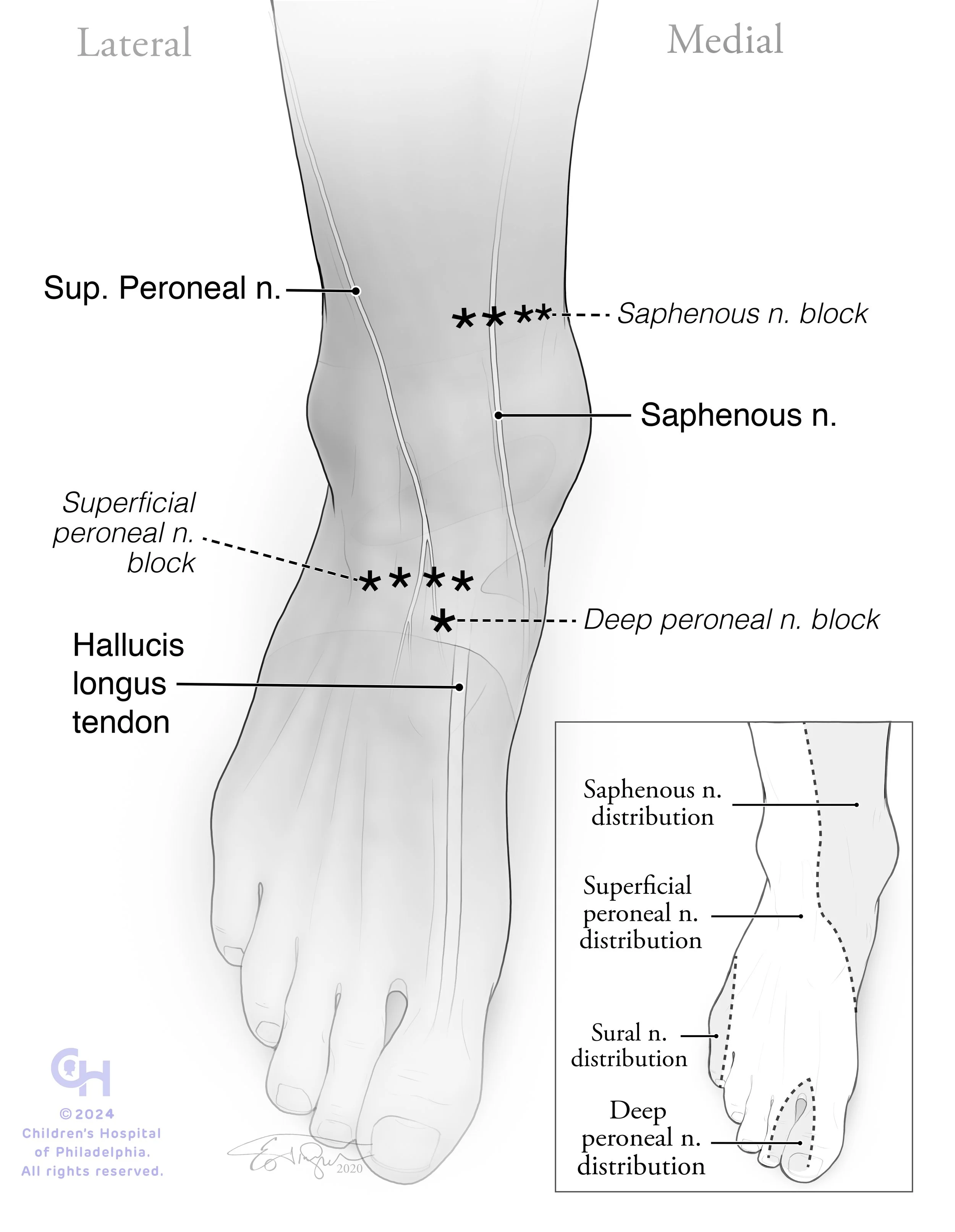
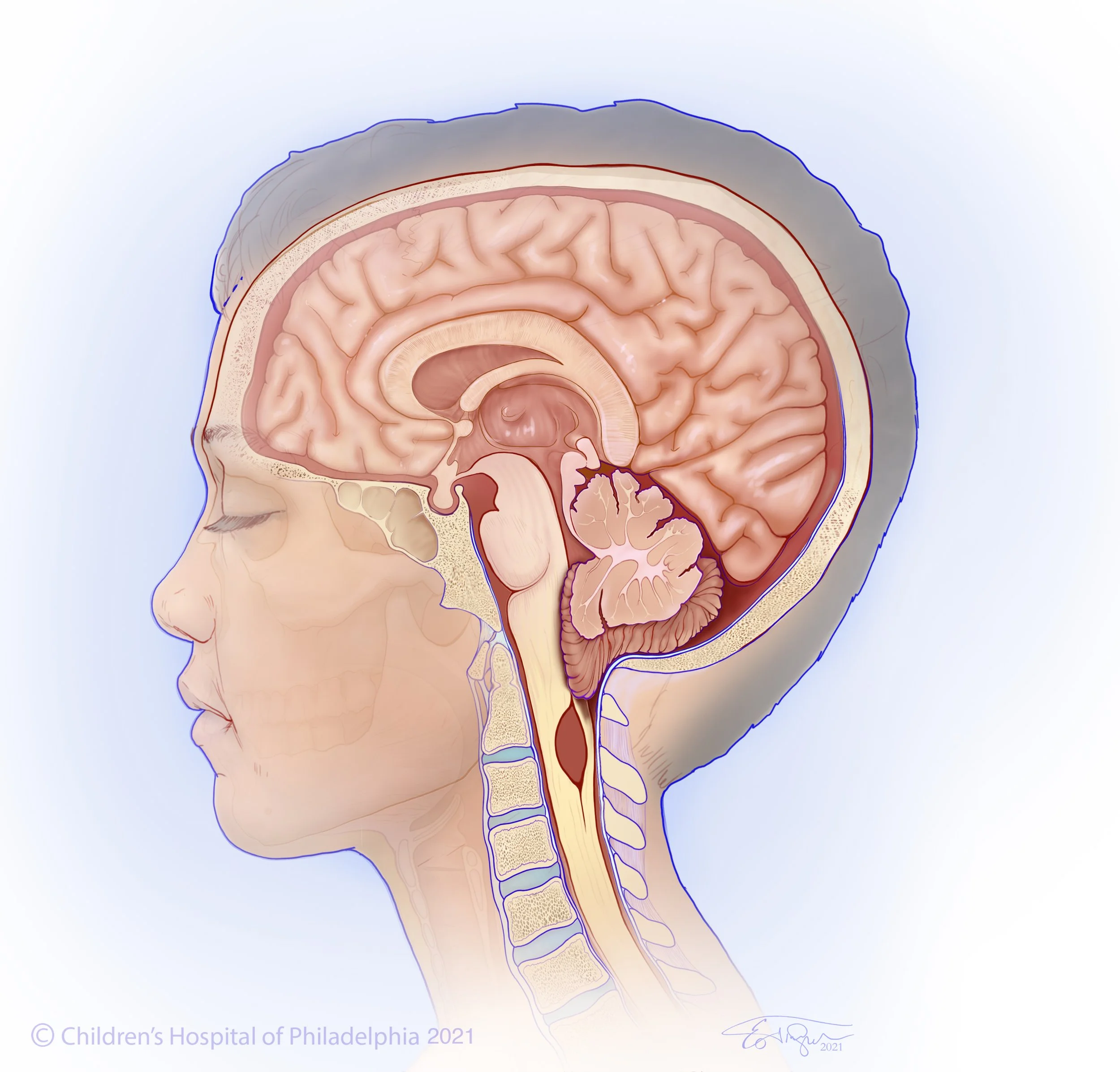
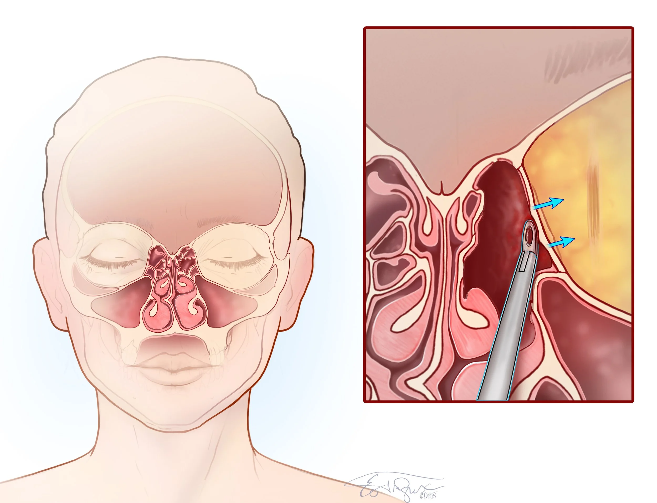
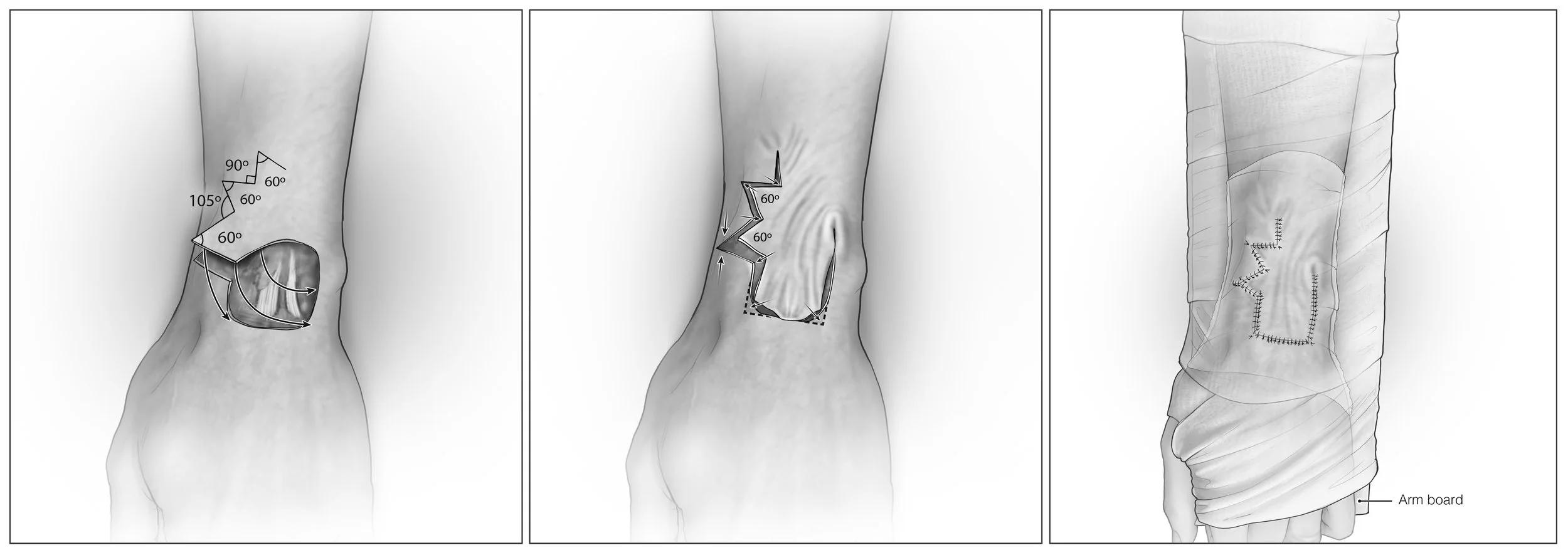
Clear, didactic illustrations and videos are cornerstones of medical education. By developing modules and curriculum wide visuals with educators, Trueblood Studio sets new standards for medical programs.















Quisque iaculis facilisis lacinia. Mauris euismod pellentesque tellus sit amet mollis.